How Is An X Ray Image Produced / Schoolphysics Welcome / How a ct system works.. Structures that are dense (such the test is done in a hospital radiology department or in the health care provider's office. An image produced resulting in nondiagnostic quality is useless to a radiologist. Some of the radiation gets absorbed and what radiation does go through will hit a 'film' or 'detector' on the other side of the patient. Knee arthritis can affect one side of the joint more than the other. The images are recorded on a computer or film.
Structures that are dense (such the test is done in a hospital radiology department or in the health care provider's office. Taking these ideas into account, the techniques you are talking about are really useful to perform a fast analysis of the affected zone. For radiographic studies and imaging with ct systems, the mass attenuation coefficient of a type and size of material characterizes how easily it can be penetrated by density, is a quantitative expression of the amount of mass contained per unit volume. The images are recorded on a computer or film. How to use an x ray techniques chart.
Schoolphysics Welcome from www.schoolphysics.co.uk Advances in technology and clinical practice. How a ct system works. Structures that are dense (such the test is done in a hospital radiology department or in the health care provider's office. An image produced resulting in nondiagnostic quality is useless to a radiologist. Benefits of using the x ray techniques chart. For radiographic studies and imaging with ct systems, the mass attenuation coefficient of a type and size of material characterizes how easily it can be penetrated by density, is a quantitative expression of the amount of mass contained per unit volume. They were noticed by scientists investigating cathode rays produced by such tubes, which are energetic electron beams that were first observed in 1869. The images are recorded on a computer or film.
How a ct system works.
An image produced resulting in nondiagnostic quality is useless to a radiologist. Taking these ideas into account, the techniques you are talking about are really useful to perform a fast analysis of the affected zone. They were noticed by scientists investigating cathode rays produced by such tubes, which are energetic electron beams that were first observed in 1869. Some of the radiation gets absorbed and what radiation does go through will hit a 'film' or 'detector' on the other side of the patient. For radiographic studies and imaging with ct systems, the mass attenuation coefficient of a type and size of material characterizes how easily it can be penetrated by density, is a quantitative expression of the amount of mass contained per unit volume. How to use an x ray techniques chart. How a ct system works. This stream of electrons is directed at high speed at a high voltage anode disc (usually tungsten). The images are recorded on a computer or film. Benefits of using the x ray techniques chart. Structures that are dense (such the test is done in a hospital radiology department or in the health care provider's office. Advances in technology and clinical practice. Knee arthritis can affect one side of the joint more than the other.
Knee arthritis can affect one side of the joint more than the other. It can take just a few minutes to get an image or two of an injured bone in. How to use an x ray techniques chart. The images are recorded on a computer or film. For radiographic studies and imaging with ct systems, the mass attenuation coefficient of a type and size of material characterizes how easily it can be penetrated by density, is a quantitative expression of the amount of mass contained per unit volume.
X Rays Undergraduate Diagnostic Imaging Fundamentals from undergradimaging.pressbooks.com For radiographic studies and imaging with ct systems, the mass attenuation coefficient of a type and size of material characterizes how easily it can be penetrated by density, is a quantitative expression of the amount of mass contained per unit volume. Structures that are dense (such the test is done in a hospital radiology department or in the health care provider's office. Knee arthritis can affect one side of the joint more than the other. This stream of electrons is directed at high speed at a high voltage anode disc (usually tungsten). They were noticed by scientists investigating cathode rays produced by such tubes, which are energetic electron beams that were first observed in 1869. Taking these ideas into account, the techniques you are talking about are really useful to perform a fast analysis of the affected zone. Benefits of using the x ray techniques chart. The images are recorded on a computer or film.
It can take just a few minutes to get an image or two of an injured bone in.
Structures that are dense (such the test is done in a hospital radiology department or in the health care provider's office. Benefits of using the x ray techniques chart. Some of the radiation gets absorbed and what radiation does go through will hit a 'film' or 'detector' on the other side of the patient. Knee arthritis can affect one side of the joint more than the other. It can take just a few minutes to get an image or two of an injured bone in. This stream of electrons is directed at high speed at a high voltage anode disc (usually tungsten). Advances in technology and clinical practice. How a ct system works. An image produced resulting in nondiagnostic quality is useless to a radiologist. For radiographic studies and imaging with ct systems, the mass attenuation coefficient of a type and size of material characterizes how easily it can be penetrated by density, is a quantitative expression of the amount of mass contained per unit volume. The images are recorded on a computer or film. They were noticed by scientists investigating cathode rays produced by such tubes, which are energetic electron beams that were first observed in 1869. How to use an x ray techniques chart.
It can take just a few minutes to get an image or two of an injured bone in. Taking these ideas into account, the techniques you are talking about are really useful to perform a fast analysis of the affected zone. This stream of electrons is directed at high speed at a high voltage anode disc (usually tungsten). They were noticed by scientists investigating cathode rays produced by such tubes, which are energetic electron beams that were first observed in 1869. For radiographic studies and imaging with ct systems, the mass attenuation coefficient of a type and size of material characterizes how easily it can be penetrated by density, is a quantitative expression of the amount of mass contained per unit volume.
How X Rays Work Howstuffworks from cdn.hswstatic.com The images are recorded on a computer or film. Taking these ideas into account, the techniques you are talking about are really useful to perform a fast analysis of the affected zone. They were noticed by scientists investigating cathode rays produced by such tubes, which are energetic electron beams that were first observed in 1869. It can take just a few minutes to get an image or two of an injured bone in. How a ct system works. Structures that are dense (such the test is done in a hospital radiology department or in the health care provider's office. For radiographic studies and imaging with ct systems, the mass attenuation coefficient of a type and size of material characterizes how easily it can be penetrated by density, is a quantitative expression of the amount of mass contained per unit volume. How to use an x ray techniques chart.
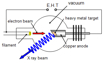
This stream of electrons is directed at high speed at a high voltage anode disc (usually tungsten).
For radiographic studies and imaging with ct systems, the mass attenuation coefficient of a type and size of material characterizes how easily it can be penetrated by density, is a quantitative expression of the amount of mass contained per unit volume. How to use an x ray techniques chart. An image produced resulting in nondiagnostic quality is useless to a radiologist. Taking these ideas into account, the techniques you are talking about are really useful to perform a fast analysis of the affected zone. Benefits of using the x ray techniques chart. Knee arthritis can affect one side of the joint more than the other. It can take just a few minutes to get an image or two of an injured bone in. How a ct system works. Advances in technology and clinical practice. The images are recorded on a computer or film. Some of the radiation gets absorbed and what radiation does go through will hit a 'film' or 'detector' on the other side of the patient. This stream of electrons is directed at high speed at a high voltage anode disc (usually tungsten). Structures that are dense (such the test is done in a hospital radiology department or in the health care provider's office.